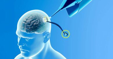
Expression of SARS-CoV-2 spike protein in cerebral Arteries: Implications for hemorrhagic stroke Post-mRNA vaccination
Source : sciencedirect.com – June 2025 – Nakao Ota, Masahiko Itani, Tomohiro Aoki, Aki Sakurai, Takashi Fujisawa, Yasuaki Okada, Kosumo Noda, Yoshiki Arakawa, Sadahisa Tokuda, Rokuya Tanikawa
https://www.sciencedirect.com/science/article/pii/S096758682500195X
Abonnez-vous au canal Telegram Strategika pour ne rien rater de notre actualité
Pour nous soutenir commandez les livres Strategika : “Globalisme et dépopulation” , « La guerre des USA contre l’Europe » et « Société ouverte contre Eurasie »
Highlights
- •Spike protein expression was detected in 43.8% of vaccinated patients.
- •SARS-CoV-2 spike protein persists in cerebral arteries up to 17 months post-vaccination.
- •Spike protein was expressed in the intima of the cerebral arteries.
- •In situ hybridization confirmed vaccine- and virus-derived spike protein mRNA.
- •Findings highlight concerns about mRNA vaccine biodistribution and long-term safety.
Abstract
Background
The rapid deployment of mRNA vaccines for SARS-CoV-2, such as BNT162b2 (BioNTech-Pfizer) and mRNA-1273 (Moderna), provided a critical tool in combating the COVID-19 pandemic. While their short-term safety and efficacy were demonstrated in clinical trials, rare adverse events, including hemorrhagic strokes, have been reported after widespread use. However, the long-term biodistribution and effects of mRNA vaccines remain underexplored.
This study aimed to investigate the long-term presence of SARS-CoV-2 spike protein in brain tissues of patients with hemorrhagic strokes, examining its potential association with mRNA vaccination.
Methods
A total of 19 cases of hemorrhagic stroke from 2023 to 2024 were retrospectively analyzed. Immunohistochemical staining for SARS-CoV-2 spike protein and nucleocapsid protein was performed on tissue samples. In situ hybridization was conducted in selected cases to confirm the origin of spike protein expression (vaccine or viral infection). Vaccination history and SARS-CoV-2 infection status were documented for all cases.
Results
Spike protein expression was detected in 43.8 % of vaccinated patients, predominantly localized to the intima of cerebral arteries, even up to 17 months post-vaccination. While no active inflammatory changes were identified, infiltration of CD4-, CD8- and CD68- positive cells was observed in the spike protein positive vessels. In situ hybridization confirmed the presence of both vaccine-derived mRNA and SARS-CoV-2 virus-derived mRNA, which encode the spike protein, in select cases. Notably, spike protein positivity was observed exclusively in female patients (P = 0.015). None of the cases showed nucleocapsid protein positivity, supporting the absence of active viral infection.
Conclusion
Although the possibility of spike protein expression due to asymptomatic SARS-CoV-2 infection cannot be entirely excluded, this study demonstrated prolonged presence of SARS-CoV-2 spike protein in the cerebral arteries following mRNA vaccination. Additionally, some inflammatory cell infiltration was observed in spike-positive vessels. These findings raise significant concerns regarding the biodistribution of lipid nanoparticle-based vaccines and their long-term safety. Global replication studies are urgently required to validate these findings and ensure comprehensive safety evaluations of mRNA vaccines.
Keywords
SARS-CoV-2 spike protein
mRNA vaccines
Hemorrhagic stroke
Spike protein expression
Lipid nanoparticle biodistribution
Long-term vaccine safety
Sex-based differences
1. Introduction
The urgent need for countermeasures against the coronavirus disease 2019 (COVID-19) pandemic has spurred the rapid development of severe acute respiratory syndrome coronavirus 2 (SARS-CoV-2) vaccines of diverse formulations. mRNA vaccines BNT162b2 (BioNTech-Pfizer) and mRNA-1273 (Moderna/NIAID) have demonstrated high efficacy and safety in clinical trials for COVID-19 prevention [1], [2], [3]. Though the emergency approval of the vaccines during the COVID-19 pandemic, humans had no prior experience with mRNA vaccines until the development of the SARS-CoV-2 mRNA vaccine, which marked the first application of this technology. As a result, compared to conventional vaccines such as inactivated or live-attenuated vaccines, there is limited experience and their long-term safety remains unknown.
The risk of stroke associated with SARS-CoV-2 infection has been reported since the early stages of the pandemic [4], [5], [6], [7]. In particular, the risk of ischemic stroke due to hypercoagulation state and endothelial injury has been highlighted [7], [8]. However, although thrombotic or hemorrhagic strokes following SARS-CoV-2 mRNA vaccination were not observed during the phase 2–3 clinical trial [3], several cases of such strokes have been reported after widespread clinical use. These reports are primarily limited to early complications occurring within approximately one month after vaccination [9], [10], [11], [12].
We encountered a case in which the SARS-CoV-2 spike protein was detected via immunohistochemical staining in brain tissue samples obtained during surgery from a patient presenting with an atypical form of cerebral hemorrhage several months after vaccination, despite no history of SARS-CoV-2 infection (Fig. 1). This observation prompted us to retrospectively and prospectively collect tissue samples from patients with hemorrhagic stroke to investigate spike protein expression and its potential association with vaccination, SARS-CoV-2 infection, and the pathogenesis of hemorrhagic stroke.

2. Materials and methods
2.1. Study design and population
From March 2023 to April 2024, we retrospectively and prospectively included all cases of patients with hemorrhagic stroke or autopsy cases who underwent surgery at Sapporo Teishinkai Hospital, with approval from the Institutional Review Board of Sapporo Teishinkai Hospital (#2023–26) and the Institutional Review Board of the Jikei University (#35–345[11977]). Written informed consent was obtained from all patients or their legal representative. During surgery, brain tissues or other samples were collected from damaged areas while removing the hematoma or repairing aneurysm. Information regarding the vaccine type and vaccination dates was retrieved from municipal vaccination records, including the dates and lot numbers (Supplemental Table 1).
One case (Case 18) involved an autopsy following death from subarachnoid hemorrhage. Vaccination records were unavailable for this case; therefore, the “time from last vaccination to surgery” was marked as NA. According to information obtained from the family during the interview, the patient had been vaccinated 15 months prior to surgery.
SARS-CoV-2 infection history was confirmed only for cases where patients had been diagnosed as positive via polymerase chain reaction (PCR) test or antigen testing at a medical institution. Therefore, cases with mild cold-like symptoms or undiagnosed infections due to the absence of medical consultation were considered as having no history of SARS-CoV-2 infection.
2.2. Histology and immunohistochemical staining
Biopsy from brain tissue, cerebral artery or other samples and autopsy tissue samples were fixed in formalin and embedded in paraffin (FFPE), and sectioned. Once unstained slides were generated and initial Hematoxylin and Eosin (H&E) stained sections were analyzed. Immunohistochemistry was performed After de-paraffinization and blocking with 10 % donkey serum (#AB_2337258, Jackson ImmunoResearch, West Grove, PA), slices were incubated with primary antibodies followed by incubation with secondary antibodies conjugated with fluorescence dye. Finally, fluorescent images were acquired on a confocal fluorescence microscope system (LSM880 or LSM980, Carl Zeiss, Oberkochen, Germany).
Primary antibodies used were as follows; rabbit monoclonal anti-Nucleocapsid antibody (Clone; HL344, #MA5-36251, Thermo Fisher Scientific, Waltham, MA), and mouse monoclonal anti-Spike Protein antibody (Clone; GT263, #MA5-36245, Thermo Fisher Scientific).
Secondary antibodies used were as follows; Alexa Fluor 488-conjugated donkey anti-mouse IgG H&L antibody (#A21202, Thermo Fisher Scientific), Alexa Fluor 647-conjugated donkey anti-rabbit IgG H&L antibody (#A31573, Thermo Fisher Scientific).
2.3. In situ hybridization
Since some cases showed positive staining for the SARS-CoV-2 spike protein via immunohistochemical staining, we planned to perform in situ hybridization to differentiate whether the spike protein expression was due to SARS-CoV-2 infection or mRNA vaccination. To address this, in situ hybridization was conducted on three cases (Case 4, 5 and 15) where the infection history was unclear, a significant amount of time had elapsed since vaccination, yet spike protein was still detected through immunostaining.
For in situ hybridization, slices from human specimens were prepared as in above. After de-paraffinization, in situ hybridization was done using a RNAscope™; Duplex Reagent Kit (#322430, Advanced Cell Diagnostics, Inc., Newark, CA) and probes to detect mRNA from vaccine (#1215391, Advanced Cell Diagnostics) and to detect mRNA encoding spike protein in SARS-CoV-2 (#848561, Advanced Cell Diagnostics) from the company. The probe to detect the gene encoding Peptidylprolyl Isomerase B (#313901, Advanced Cell Diagnostics) was used as a positive control experiment and one to detect bacterial gene for Dihydrodipicolinate Reductase (dapB) from Bacillus subtilis strain SMY (#310043, Advanced Cell Diagnostics) as a negative control experiment. The images were acquired by a microscopic system.
The negative control for in situ hybridization was obtained from Case 15.
2.4. Statistical Analysis
Data are presented as the mean ± standard deviation, median (interquartile range), or number of patients (%), as appropriate. Fisher’s exact test was employed to analyze the association between spike protein positivity and sex differences. Statistical analyses were performed using IBM SPSS Statistics for Mac, version 27.0 (IBM Corp., Armonk, NY, USA). A P-value of < 0.05 was considered statistically significant.
3. Results
Nineteen cases were included in the study, with details summarized in Table 1. Of these, 10 were intracerebral hemorrhage, subarachnoid hemorrhage in 6 cases, hemorrhage due to arterio-venous malformation in 2 and one case hemorrhage due to cavernous malformation. The mean age was 58.9 (±18.9) years, with a predominance of females (14 cases, 73.7 %). Sixteen patients had received an mRNA vaccine, while three had not. Among the 16 vaccinated patients, the median number of vaccinations was 4.0 (IQR 2.0), and the mean time from the last vaccination to surgery was 10.8(±6.8) months.
Table 1. Study Cases: Baseline Characteristics, Immunohistochemical Staining Results, and Histories of Vaccination and COVID-19 Infection.
| Case | Age | SEX | Diagnosis | Sample | History of previous diagnosis of COVID19 infection | History of mRNA Vaccinations | Number of Vaccinations | Time from last vaccination to surgery (month) | Immunostaining Spike protein/Nucleocapsid |
|---|---|---|---|---|---|---|---|---|---|
| 1 | 25 | Female | Subcortical hemorrhage | AVM | NO | YES | 3 | 5 | Negative/ Negative |
| 2 | 75 | Male | Putaminal hemorrhage | Brain tissue | NO | YES | 4 | 7 | Negative/ Negative |
| 3 | 50 | Female | Putaminal hemorrhage | Brain tissue | NO | YES | 2 | 22 | Negative/ Negative |
| 4* | 70 | Female | Subarachnoid hemorrhage | Cerebral artery | NO | YES | 3 | 17 | Positive/ Negative |
| 5* | 79 | Female | Cerebellar hemorrhage | Brain tissue | NO | YES | 5 | 12 | Positive/ Negative |
| 6 | 49 | Female | Putaminal hemorrhage | Brain tissue | NO | NO | 0 | − | Positive/ Negative |
| 7 | 53 | Male | Cerebellar hemorrhage | Brain tissue | NO | YES | 3 | 18 | Negative/ Negative |
| 8 | 63 | Male | Putaminal hemorrhage | Brain tissue | NO | NO | 0 | − | Negative/ Negative |
| 9 | 72 | Female | Subcortical hemorrhage | Brain tissue | NO | YES | 7 | 5 | NA |
| 10 | 23 | Female | Intraventricular hemorrhage | AVM | YES | NO | 0 | − | Positive/ Negative |
| 11 | 67 | Male | Putaminal hemorrhage | Brain tissue | NO | YES | 4 | 2 | Negative/ Negative |
| 12 | 81 | Female | Subarachnoid hemorrhage | Brain tissue | NO | YES | 7 | 2 | Positive/ Negative |
| 13 | 50 | Female | Subarachnoid hemorrhage | Cervical lymph node | NO | YES | 5 | 3 | Negative/ Negative |
| 14 | 55 | Female | Subarachnoid hemorrhage | Brain tissue | NO | YES | 3 | 21 | Negative/ Negative |
| 15* | 71 | Female | Putaminal hemorrhage | Brain tissue | NO | YES | 4 | 11 | Positive/ Negative |
| 16 | 88 | Female | Subcortical hemorrhage | Brain tissue | NO | YES | 7 | 8 | Positive/ Negative |
| 17 | 47 | Male | Brain stem hemorrhage | Cavernous malformation | YES | YES | 4 | 15 | Negative/ Negative |
| 18 | 72 | Female | Subarachnoid hemorrhage | Brain tissue | NO | YES | 3 | NA | Positive/ Negative |
| 19 | 30 | Female | Subarachnoid hemorrhage | Brain tissue | NO | YES | 5 | 14 | Positive/ Negative |
AVM; arterio-venous malformation, *; patients with being performed in situ hybridization
3.1. Immunohistochemical staining for spike protein and nucleocapsid protein
Fig. 1 shows an illustrative case in which SARS-CoV-2 spike protein was detected via immunohistochemical staining in brain tissue samples (Case 9). A 72-year-old woman with a history of seven SARS-CoV-2 vaccinations and no history of COVID-19 infection underwent emergent clot removal surgery for an intracerebral hemorrhage in the left temporal lobe. Specimens were collected from the area surrounding the hemorrhage. Based on the immunohistochemical staining results, cerebral amyloid angiopathy was ruled out, and positive staining for the spike protein was observed in the intima of the vessel and smooth muscle.
Fig. 2 shows the results of immunostaining for SARS-CoV-2 spike protein and nucleocapsid protein. Among the 16 patients with a history of mRNA vaccination, 7 cases (43.8 %) showed a positive staining for the SARS-CoV-2 spike protein (Cases 4, 5, 6, 10, 12, 15, 16, 18, and 19), with no positive staining for the nucleocapsid protein. Among the 3 patients without a history of mRNA vaccination (Case 6, 8 and 10), 2 cases (66.7 %) were positive staining for the SARS-CoV-2 spike protein (Case 6 and 10) and negative staining for nucleocapsid protein. The remaining patient negative staining for the nucleocapsid protein but had a history of a SARS-CoV-2 infection (Case 8).

In the positive staining of the spike protein cases, all spike protein was expressed in the intima of the cerebral arteries. No obvious aggressive inflammatory cells infiltration suggesting active vasculitis was observed. However, in the spike protein-positive cases, whether with or without history of vaccination, findings such as CD4-positive T-cell and CD68-positive cells infiltration into the intima, as well as CD 8-positive cells infiltration into the adventitia, were noted. In the spike protein negative cases, no such findings were observed (Fig. 3, Fig. 4).


Among the 18 cases, excluding the case of defective specimen, all cases with positive spike protein staining (9 females and 0 males) were female (Fisher’s exact test; P = 0.015) (Table 1).
3.2. In situ hybridization
In situ hybridization was conducted using control probes, SARS-CoV-2 vaccine mRNA-specific probes, and spike protein mRNA probes for Cases 4, 5, and 15. These cases showed positive staining for the SARS-CoV-2 spike protein and negative for the nucleocapsid protein in immunohistochemical staining. It had been 17, 12, and 11 months since their last mRNA-1273 or BNT162b2 vaccination. In all three cases, in situ hybridization revealed the presence of both mRNA from the vaccine and SARS-CoV-2 viral RNA in the intima (Fig. 5).

4. Discussion
The present study revealed that spike protein derived from the mRNA vaccine was expressed in the intima of the cerebral arteries in patients with hemorrhagic stroke, even 17 months after vaccination. The relationship between hemorrhagic stroke and the expression of spike protein remains unclear, as the excised samples were not from hemorrhagic lesions themselves, and there was no obvious evidence of active vasculitis, but showed infiltration of some inflammatory cells in the spike protein-positive vessels. To the best of our knowledge, this is the first report of its kind worldwide.
There are reports of hemorrhagic stroke complications occurring in the acute stage, within one month after vaccination. These reports are primarily based on circumstantial evidence of atypical strokes shortly following vaccination [9], [10], [12]. A large epidemiologic study in Hong Kong also demonstrated that the BNT162b2 (Pfizer-BioNTech) vaccine was associated with an increased risk of hemorrhagic stroke during the 14–27 days after the first dose and the 0–13 days after the second dose. However, the study also concluded that the incidence of thromboembolic events and hemorrhagic stroke was lower among vaccine recipients compared to individuals who tested positive for SARS-CoV-2, affirming the vaccine’s safety [10]. To date, there have been no reports of long-term complications following SARS-CoV-2 vaccination.
The reported mechanism of the ischemic and hemorrhagic stroke due to spike protein of SARS-CoV-2 are that, the SARS-CoV-2 spike protein has been implicated in disrupting blood–brain barrier integrity through RhoA activation, potentially contributing to neurological complications in COVID-19 patients [4]. The virus’s binding to angiotensin converting enzyme-2 (ACE2) receptors can lead to abnormal blood pressure regulation, increasing the risk of hemorrhagic stroke [7]. COVID-19-associated coagulopathy, characterized by elevated D-dimer and inflammatory markers, may predispose patients to both ischemic and hemorrhagic strokes [5], [6]. The infection can induce a hypercoagulable state, disrupt endothelial function, and trigger an inflammatory response, collectively increasing stroke risk [6].
Although our study did not reveal infiltration of inflammatory cells suggesting active vasculitis in the intima, findings such as CD4-positive T-cell and CD68-positive cells infiltration into the intima, as well as CD8-positive cells infiltration into the adventitia, were observed. This may be because the specimen was taken from the surrounding brain tissues rather than the hemorrhagic point itself. Therefore, we hypothesize that expression of the spike protein drive T cell immunity, leading to the hemorrhagic stroke.
Recent studies have explored the biodistribution of SARS-CoV-2 mRNA vaccines in humans. Vaccine-associated mRNA has been detected in blood for at least 15 days post-vaccination [13], with some studies reporting its presence up to 28 days [14]. However, the biodistribution of SARS-CoV-2 mRNA vaccines in body tissues remains poorly understood. Lipid nanoparticles, which encapsulate the mRNA, may cross physiological barriers such as the blood–brain barrier, blood-placental barrier [15]. Additionally, recent research has examined the biodistribution and transferability of SARS-CoV-2 vaccines to human tissues. Concerns have been raised regarding potential autoimmune inflammatory reactions in terminally differentiated tissues like the heart and brain, triggered by in situ spike protein production [16], [17]. Case reports have documented multifocal necrotizing encephalitis and myocarditis [16] as well as fatal multi-organ inflammation following SARS-CoV-2 vaccination [18]. These adverse effects were predominantly reported within a month after vaccination.
The present study revealed that lipid nanoparticle-modified mRNA vaccines could induce spike protein production in the vessel walls of the brain, persisting longer than previously anticipated. The actual behavior of mRNA vaccines within the human body may differ from what was previously believed. We suggest, based on the results of this study, that global replication studies should be conducted to verify the true safety profile of mRNA vaccines. These efforts are necessary to address potential concerns and ensure a comprehensive understanding of their long-term effects.
Recent research highlights significant sex differences in both acute COVID-19 and post-acute sequelae of SARS-CoV-2 infection. During the acute phase, men tend to exhibit higher levels of innate immune cytokines and experience more severe symptoms, whereas women demonstrate stronger T cell activation [19]. In the long term, women are more likely to report persistent symptoms, such as dyspnea, fatigue, chest pain, and palpitations [20]. Studies have also shown that women are more likely to experience psychiatric or mood disorders, ear, nose, and throat issues, musculoskeletal problems, and neurological conditions as part of long COVID syndrome [21].
Although based on a small number of cases, the findings of the present study—particularly the significant sex-based differences in spike protein expression within the intima of cerebral arteries—may provide insights into the underlying mechanisms contributing to these observed disparities between sexes.
5. Limitation of the study
In our study, in situ hybridization detected both mRNA derived from the vaccine and mRNA from the SARS-CoV-2 virus. This suggests that the observed spike protein positive staining in patients with a history of SARS-CoV-2 vaccination, but no documented history of viral infection, cannot definitively be attributed to the vaccine alone. Notably, the nucleocapsid protein was consistently negative across cases, supporting the notion that our in situ hybridization method has high sensitivity and could detect trace amounts of mRNA, possibly reflecting unrecognized asymptomatic infections. These findings emphasize the need for caution in interpreting the presence of spike protein as exclusively vaccine-related.
Another limitation of this study is small sample size. It is necessary to expand the number of cases and clarify the true incidence and epidemiology. However, the fact that more than half of the cases in the present study express the spike protein in brain tissue is quite important. Based on these findings, global replication studies should be conducted to verify the true safety profile of mRNA vaccines.
6. Conclusions
Although the possibility of spike protein expression due to asymptomatic SARS-CoV-2 infection cannot be entirely excluded, this study demonstrated that mRNA vaccines for SARS-CoV-2 can result in prolonged spike protein expression within the intima of cerebral arteries, persisting for months after vaccination. This raises significant concerns regarding the biodistribution of lipid nanoparticle-encapsulated mRNA and the potential for long-term, yet rare, adverse events. Additionally, the notable presence of spike protein in vaccinated individuals and some inflammatory cell infiltration was observed in spike-positive vessels—particularly women—highlights potential sex-based differences in vaccine response. These results underscore the urgent global studies to replicate and expand upon these findings, ensuring a more comprehensive understanding of the long-term safety profile of mRNA vaccines.
Further research is also needed to differentiate between spike protein derived from vaccination and that resulting from undiagnosed asymptomatic SARS-CoV-2 infection. Improved detection methods and robust epidemiological studies could mitigate current limitations and better inform clinical and public health strategies. Ultimately, while mRNA vaccines remain a critical tool against COVID-19, ongoing evaluation of their long-term effects is essential for ensuring their safety and effectiveness.
Sources of Funding
This work was supported by Teishinkai Foundation.
CRediT authorship contribution statement
Nakao Ota: Writing – original draft, Resources, Project administration, Methodology, Investigation, Conceptualization. Masahiko Itani: Writing – review & editing, Validation, Investigation, Data curation. Tomohiro Aoki: Writing – review & editing, Validation, Methodology, Investigation, Data curation, Conceptualization. Aki Sakurai: Writing – review & editing, Supervision, Methodology. Takashi Fujisawa: Writing – review & editing, Resources, Data curation. Yasuaki Okada: Writing – review & editing, Resources, Data curation. Kosumo Noda: Writing – review & editing, Resources. Yoshiki Arakawa: Writing – review & editing, Supervision. Sadahisa Tokuda: Writing – review & editing, Funding acquisition. Rokuya Tanikawa: Writing – review & editing, Supervision, Conceptualization.
Declaration of competing interest
The authors declare that they have no known competing financial interests or personal relationships that could have appeared to influence the work reported in this paper.
Acknowledgements
We thank Shota Watanabe, Pharmacist at Sapporo Teishinkai Hospital, for being responsible for collecting the necessary data.
Appendix A. Supplementary data
The following are the Supplementary data to this article:
Download: Download Acrobat PDF file (81KB)
Supplementary Data 1.
References
- [1]L.R. Baden, H.M. El Sahly, B. Essink, K. Kotloff, S. Frey, R. Novak, et al.Efficacy and Safety of the mRNA-1273 SARS-CoV-2 VaccineN Engl J Med, 384 (2021), pp. 403-416, 10.1056/NEJMoa2035389
View at publisherView in ScopusGoogle Scholar[2]
F.P. Polack, S.J. Thomas, N. Kitchin, J. Absalon, A. Gurtman, S. Lockhart, et al.
Safety and Efficacy of the BNT162b2 mRNA Covid-19 Vaccine
N Engl J Med, 383 (2020), pp. 2603-2615, 10.1056/NEJMoa2034577View at publisherView in ScopusGoogle Scholar[3]
S.J. Thomas, E.D. Moreira Jr., N. Kitchin, J. Absalon, A. Gurtman, S. Lockhart, et al.
Safety and Efficacy of the BNT162b2 mRNA Covid-19 Vaccine through 6 Months
N Engl J Med, 385 (2021), pp. 1761-1773, 10.1056/NEJMoa2110345View at publisherGoogle Scholar[4]
B.J. DeOre, K.A. Tran, A.M. Andrews, S.H. Ramirez, P.A. Galie
SARS-CoV-2 Spike Protein Disrupts Blood-Brain Barrier Integrity via RhoA Activation
J Neuroimmune Pharmacol, 16 (2021), pp. 722-728, 10.1007/s11481-021-10029-0View in ScopusGoogle Scholar[5]
A.A. Divani, S. Andalib, M. Di Napoli, S. Lattanzi, M.S. Hussain, J. Biller, et al.
Coronavirus Disease 2019 and Stroke: Clinical Manifestations and Pathophysiological Insights
J Stroke Cerebrovasc Dis, 29 (2020), Article 104941, 10.1016/j.jstrokecerebrovasdis.2020.104941View PDFView articleView in ScopusGoogle Scholar[6]
X. Qi, K.A. Keith, J.H. Huang
COVID-19 and stroke: A review
Brain Hemorrhages, 2 (2021), pp. 76-83, 10.1016/j.hest.2020.11.001View PDFView articleGoogle Scholar[7]
Z. Wang, Y. Yang, X. Liang, B. Gao, M. Liu, W. Li, et al.
COVID-19 Associated Ischemic Stroke and Hemorrhagic Stroke: Incidence, Potential Pathological Mechanism, and Management
Front Neurol, 11 (2020), Article 571996, 10.3389/fneur.2020.571996View in ScopusGoogle Scholar[8]
H.S. Markus, M. Brainin
COVID-19 and stroke-A global World Stroke Organization perspective
Int J Stroke, 15 (2020), pp. 361-364, 10.1177/1747493020923472View in ScopusGoogle Scholar[9]
M. Ancau, F. Liesche-Starnecker, J. Niederschweiberer, S.M. Krieg, C. Zimmer, C. Lingg, et al.
Case Series: Acute Hemorrhagic Encephalomyelitis After SARS-CoV-2 Vaccination
Front Neurol, 12 (2021), Article 820049, 10.3389/fneur.2021.820049
C.S.L. Chui, M. Fan, E.Y.F. Wan, M.T.Y. Leung, E. Cheung, V.K.C. Yan, et al.
Thromboembolic events and hemorrhagic stroke after mRNA (BNT162b2) and inactivated (CoronaVac) covid-19 vaccination: A self-controlled case series study
EClinicalMedicine, 50 (2022), Article 101504, 10.1016/j.eclinm.2022.101504View PDFView articleView in ScopusGoogle Scholar[11]
M. Sharifian-Dorche, M. Bahmanyar, A. Sharifian-Dorche, P. Mohammadi, M. Nomovi, A. Mowla
Vaccine-induced immune thrombotic thrombocytopenia and cerebral venous sinus thrombosis post COVID-19 vaccination; a systematic review
J Neurol Sci, 428 (2021), Article 117607, 10.1016/j.jns.2021.117607View PDFView articleView in ScopusGoogle Scholar[12]
R. Takeyama, K. Fukuda, Y. Kouzaki, T. Koga, S. Hayashi, H. Ohtani, et al.
Intracerebral hemorrhage due to vasculitis following COVID-19 vaccination: a case report
Acta Neurochir (Wien), 164 (2022), pp. 543-547, 10.1007/s00701-021-05038-0View in ScopusGoogle Scholar[13]
T.E. Fertig, L. Chitoiu, D.S. Marta, V.S. Ionescu, V.B. Cismasiu, E. Radu, et al.
Vaccine mRNA Can Be Detected in Blood at 15 Days Post-Vaccination
Biomedicines, 10 (2022), 10.3390/biomedicines10071538
J.A.S. Castruita, U.V. Schneider, S. Mollerup, T.D. Leineweber, N. Weis, J. Bukh, et al.
SARS-CoV-2 spike mRNA vaccine sequences circulate in blood up to 28 days after COVID-19 vaccination
APMIS, 131 (2023), pp. 128-132, 10.1111/apm.13294View in ScopusGoogle Scholar[15]
I. Pateev, K. Seregina, R. Ivanov, V. Reshetnikov
Biodistribution of RNA Vaccines and of Their Products: Evidence from Human and Animal Studies
Biomedicines, 12 (2023), 10.3390/biomedicines12010059
M. Morz
A Case Report: Multifocal Necrotizing Encephalitis and Myocarditis after BNT162b2 mRNA Vaccination against COVID-19
Vaccines (Basel), 10 (2022), 10.3390/vaccines10101651
P. Polykretis, A. Donzelli, J.C. Lindsay, D. Wiseman, A.M. Kyriakopoulos, M. Morz, et al.
Autoimmune inflammatory reactions triggered by the COVID-19 genetic vaccines in terminally differentiated tissues
Autoimmunity, 56 (2023), Article 2259123, 10.1080/08916934.2023.2259123View in ScopusGoogle Scholar[18]
H. Nushida, A. Ito, H. Kurata, H. Umemoto, I. Tokunaga, H. Iseki, et al.
A case of fatal multi-organ inflammation following COVID-19 vaccination
Leg Med (Tokyo), 63 (2023), Article 102244, 10.1016/j.legalmed.2023.102244View PDFView articleView in ScopusGoogle Scholar[19]
T. Takahashi, M.K. Ellingson, P. Wong, B. Israelow, C. Lucas, J. Klein, et al.
Sex differences in immune responses that underlie COVID-19 disease outcomes
Nature, 588 (2020), pp. 315-320, 10.1038/s41586-020-2700-3
View at publisherView in ScopusGoogle Scholar[20]
G. Pela, M. Goldoni, E. Solinas, C. Cavalli, S. Tagliaferri, S. Ranzieri, et al.
Sex-Related Differences in Long-COVID-19 Syndrome
J Womens Health (Larchmt), 31 (2022), pp. 620-630, 10.1089/jwh.2021.0411View at publisherView in ScopusGoogle Scholar[21]
S.V. Sylvester, R. Rusu, B. Chan, M. Bellows, C. O’Keefe, S. Nicholson
Sex differences in sequelae from COVID-19 infection and in long COVID syndrome: a review
Curr Med Res Opin, 38 (2022), pp. 1391-1399, 10.1080/03007995.2022.2081454